Opis
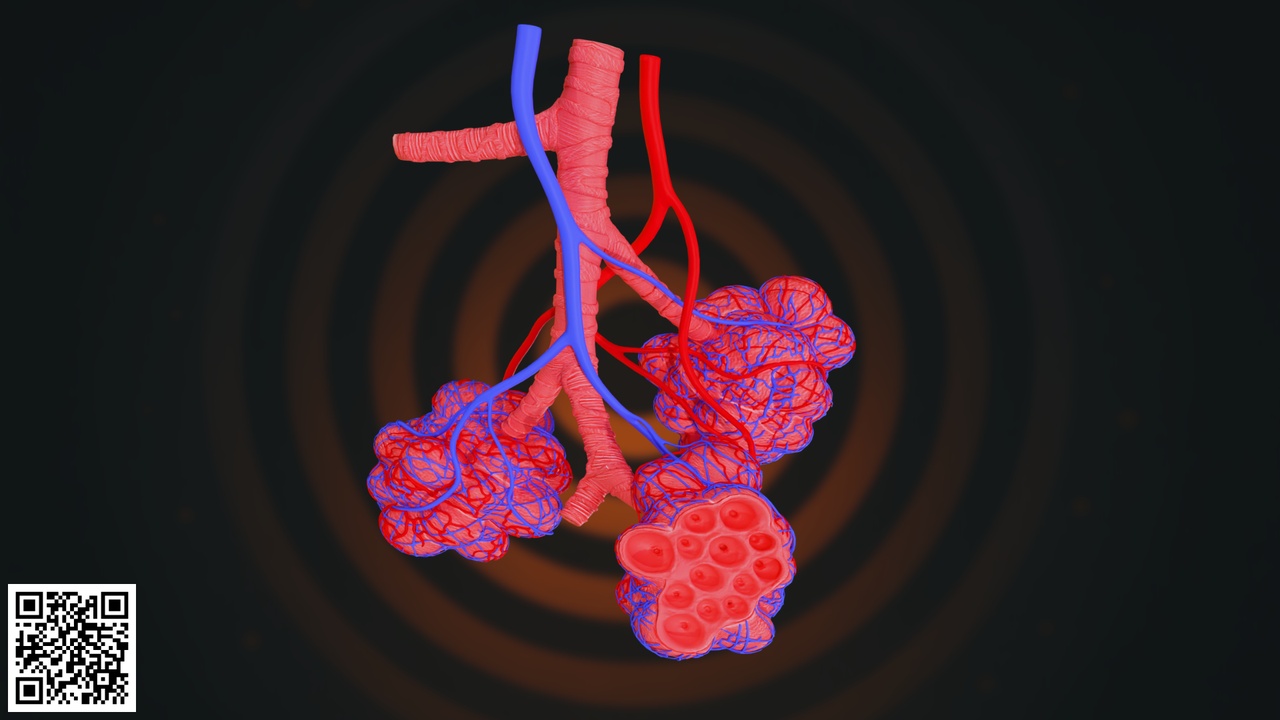
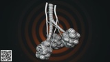
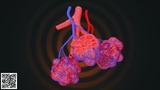
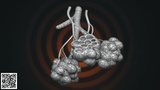
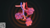
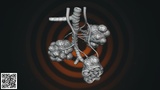
This highly detailed and anatomically accurate Human Pulmonary Alveoli 3D Model represents the microscopic gas exchange units of the lungs in a clear and educational format. The model illustrates the terminal bronchiole branching into clusters of alveolar sacs surrounded by an intricate capillary network.
The structure has been carefully designed to demonstrate the relationship between the respiratory airways and the surrounding blood vessels responsible for oxygen and carbon dioxide exchange.
The model clearly displays:
Terminal bronchiole
Alveolar ducts
Alveolar sacs
Individual alveoli
Pulmonary arteries
Pulmonary veins
Dense capillary network surrounding alveoli
Special attention has been given to anatomical accuracy, structural branching, and vascular integration. The red and blue vascular elements visually distinguish oxygenated and deoxygenated blood flow, making the model highly effective for educational and presentation purposes.
The cross-sectional alveolar detail highlights the thin-walled structure essential for efficient gas diffusion. The dense capillary wrapping demonstrates the close contact between air and blood necessary for respiratory exchange.
The mesh structure is clean and well-organized, allowing easy modification, material adjustments, and integration into larger respiratory system projects. The model is optimized for high-resolution rendering as well as real-time medical visualization workflows.
This asset is ideal for:
Medical and nursing education
Pulmonology training
Biology and anatomy classes
Healthcare presentations
Scientific visualization projects
AR/VR medical simulations
Educational animations and documentaries
Key Features:
Accurate alveolar microanatomy
Detailed capillary network
Visible bronchial branching
Color-coded vascular system
Clean topology and organized mesh
Professional educational asset