설명
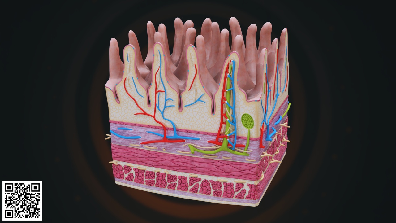
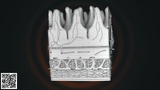
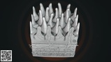
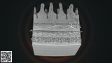
This highly detailed and anatomically accurate Human Small Intestine Wall 3D Model illustrates the microscopic structure of the intestinal lining, focusing on the finger-like projections known as villi that play a crucial role in nutrient absorption.
The model presents a cross-sectional view of the intestinal wall, revealing multiple tissue layers including the mucosa, submucosa, muscularis externa, and underlying connective tissue. The inner surface is lined with numerous villi extending into the lumen, significantly increasing the surface area for efficient digestion and absorption.
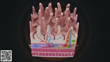
Each villus is carefully designed to demonstrate its internal components, including:
Blood capillary network (arteries and veins)
Central lacteal (lymphatic vessel)
Connective tissue core
Epithelial lining
Goblet cells (mucus-secreting cells)
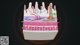
The red and blue vascular structures represent oxygenated and deoxygenated blood flow, while the green lymphatic structure highlights fat absorption through the lacteal system. The layered muscle structure beneath the mucosa shows circular and longitudinal muscle fibers responsible for peristalsis.
Special attention has been given to anatomical layering, microvascular organization, and structural depth to provide a clear educational representation of intestinal absorption mechanisms.
This model is ideal for:
Medical and nursing education
Gastroenterology training
Biology and anatomy classes
Digestive system demonstrations
Healthcare presentations
Scientific visualization projects
AR/VR educational simulations
Key Features:
Detailed villi structure
Visible blood and lymphatic supply
Multi-layer intestinal wall representation
Educational color-coded anatomy
Clean and organized mesh topology
High-resolution rendering ready