Description
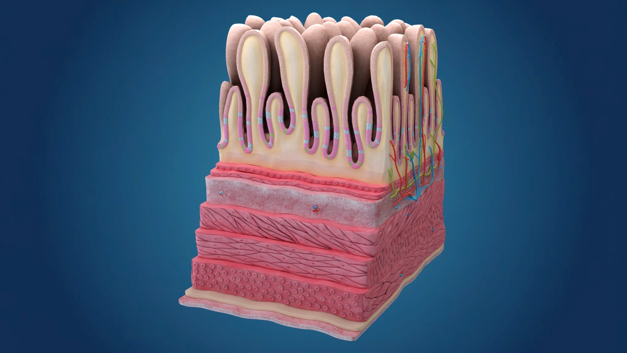
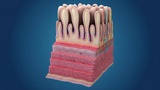
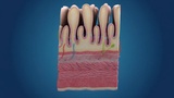
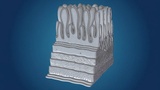
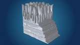
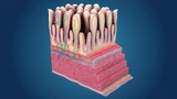
This is a high-quality, scientifically accurate 3D cross-section model of Human Intestinal Villi. This model illustrates the complex inner structure of the small intestine, specifically designed to show how the body absorbs nutrients. It features a detailed representation of the finger-like projections (villi), the epithelial layer, and the internal vascular system.
Detailed Anatomical FeaturesVilli Structure: Realistic finger-like projections that increase surface area for absorption.
Vascular Network: Highly detailed capillary network (Arteries in red, Veins in blue) for nutrient transport.
Lacteal System: Includes the lymphatic vessel (Lacteal) shown in green, responsible for fat absorption.
Tissue Layers: Clear visualization of the mucosal and sub-mucosal layers, including muscle fibers and cellular structures.
Software: Created with precision in Blender.
Formats: The package includes the original .blend file (with materials) and a universal .fbx file for seamless integration into other 3D software like Unity, Unreal Engine, or Maya.
Geometry: Clean topology, suitable for both high-end renders and real-time educational applications.
Texturing: Professionally textured to highlight different biological components clearly.
Medical Animations: Perfect for explaining digestion and nutrient absorption.
VR/AR Education: High fidelity for immersive medical training and biology apps.
Textbooks & Presentations: A great visual aid for scientific publishing and classroom learning.