
설명
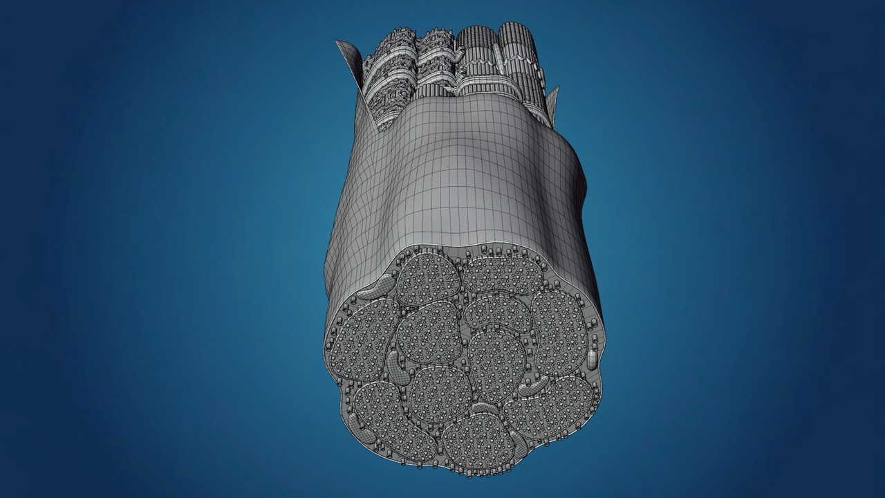
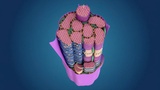
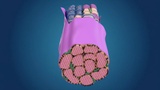
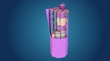
This is a high-quality, scientifically accurate 3D model of a Skeletal Muscle Fiber, showcasing the complex internal organization of a myofibril. This model is specifically designed to illustrate the microscopic anatomy of muscle tissue, making it an essential asset for medical education, physiological research, and high-end scientific visualization.
Detailed Anatomical FeaturesMyofibril Structure: Detailed representation of the rod-like units that make up a muscle fiber.
Sarcomere Units: Clearly shows the segments of the myofibril responsible for muscle contraction.
Sarcoplasmic Reticulum: Features the blue-colored network that stores and releases calcium ions.
T-Tubules: Accurately placed transverse tubules that conduct nerve impulses.
Mitochondria: Highly detailed organelles shown within the fiber to represent energy production.
Sarcolemma: The outer protective sheath of the muscle fiber, partially cut away to reveal internal details.
Blender (.blend): The original source file with procedural materials, organized layers, and optimized lighting setups.
FBX (.fbx): A high-performance, universal file format compatible with all major 3D software including Unity, Unreal Engine, Maya, and 3ds Max.
Geometry: Clean and optimized mesh topology, perfect for both close-up high-resolution renders and real-time educational applications.
Medical Training: Perfect for explaining muscle contraction mechanisms ($Actin$ and $Myosin$ interaction).
Scientific CGI: Great for documentaries, healthcare presentations, and textbook illustrations.
Interactive Learning: Ready for integration into VR/AR biology and sports science app