설명
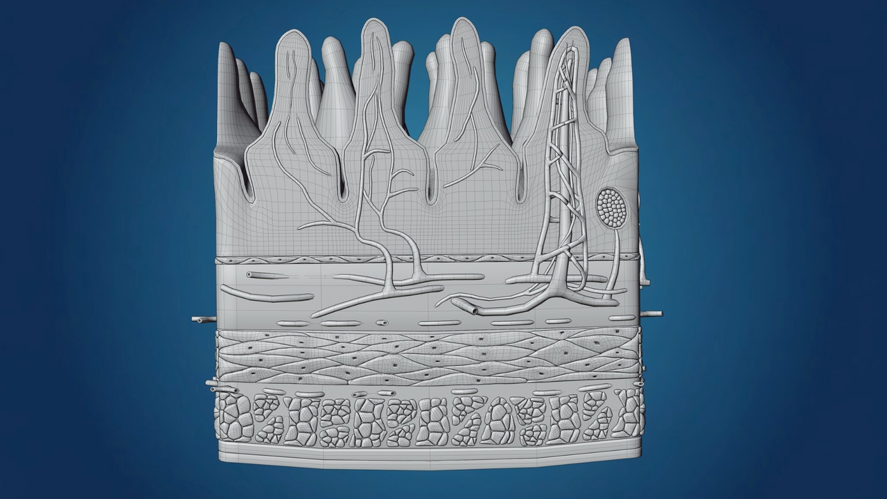
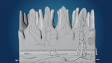
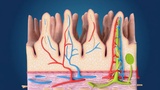
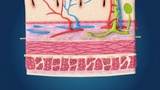
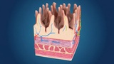
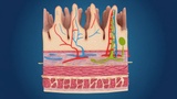
This 3D model is a highly detailed, scientifically accurate representation of the Human Small Intestinal Villi. These microscopic, finger-like projections are crucial for the digestive system as they significantly increase the surface area for nutrient absorption. This model provides a clear look into the internal and external structure of the intestinal lining.
Shutterstock
Detailed Biological FeaturesVilli Projections: Individually modeled finger-like structures that demonstrate the lining of the small intestine.
Vascular Network: Shows the intricate Capillary Loops (Arteries in red and Veins in blue) within each villus for direct blood absorption.
Lacteal System: Includes the lymphatic vessel (Lacteal), typically shown in green, which is responsible for absorbing fats and fat-soluble vitamins.
Tissue Layers: Visualization of the epithelial cells, goblet cells, and the underlying connective tissue (lamina propria).
Blender (.blend): The original source file with procedural materials and organized hierarchy.
FBX (.fbx): A high-performance, universal file format compatible with Unity, Unreal Engine, Maya, and 3ds Max.
Topology: Clean, high-resolution mesh optimized for medical animations and close-up renders.
Medical Education: Perfect for university-level biology and gastroenterology lectures.
Healthcare VR/AR: Optimized for interactive healthcare simulations.
Scientific Illustration: High-quality geometry for rendering professional-grade scientific posters or textbook diagrams.