
Beschreibung
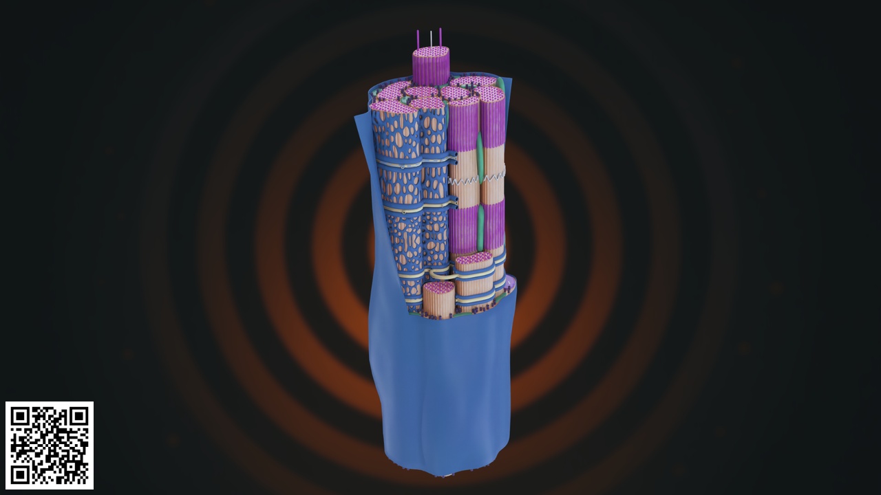
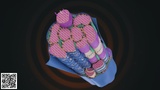
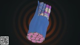
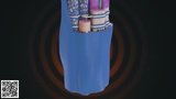
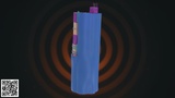
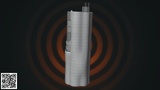
This highly detailed and anatomically accurate Human Skeletal Muscle Fiber 3D Model represents the microscopic structure of voluntary muscle tissue. The model illustrates a longitudinal and cross-sectional view of a single muscle fiber, revealing the internal organization of myofibrils and sarcomeres responsible for muscle contraction.
The outer blue layer represents the sarcolemma, enclosing the cylindrical muscle fiber. Inside, multiple tightly packed myofibrils are arranged in parallel bundles, demonstrating the contractile units that generate force. Each myofibril is segmented into repeating sarcomere units, highlighted by alternating band patterns that reflect the structural alignment of actin and myosin filaments.
The model clearly visualizes:
Muscle fiber (muscle cell)
Sarcolemma (outer membrane)
Myofibrils
Sarcomere structure
A bands and I bands
Z lines
Actin filaments
Myosin filaments
Transverse tubules (T-tubules)
Sarcoplasmic reticulum
Special attention has been given to structural accuracy, cylindrical organization, and internal layering. The cross-sectional portion reveals the dense packing of myofibrils within the sarcoplasm, making the model ideal for microscopic anatomy visualization.
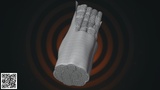
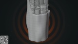
The mesh topology is clean and optimized for rendering, educational presentations, medical simulations, and animation workflows. Materials are color-coded to distinguish contractile filaments and structural components for enhanced clarity.
This model is ideal for:
Medical and nursing education
Physiology classes
Sports science training
Muscle contraction demonstrations
Healthcare presentations
AR/VR medical simulations
Scientific animation projects
Key Features:
Detailed sarcomere segmentation
Clear longitudinal and cross-section view
Accurate contractile filament arrangement
Educational color coding
Clean topology and organized mesh
High-resolution presentation ready