

Beschreibung
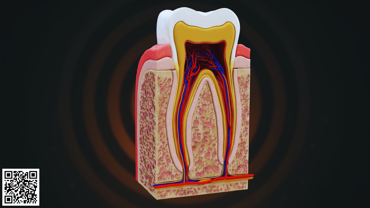
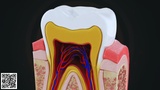
This highly detailed and anatomically accurate Human Tooth Cross-Section 3D Model presents a comprehensive internal view of tooth structure, clearly illustrating its protective outer layers and vital internal components.
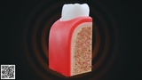
The model displays a vertical section of a human tooth embedded within the surrounding gingiva and alveolar bone. The crown portion is covered by enamel, the hardest substance in the human body, designed to protect the tooth from mechanical and chemical damage. Beneath the enamel lies the dentin, a calcified tissue that supports the crown and transmits sensory signals.
At the core of the tooth is the pulp cavity, which contains:
Dental pulp tissue
Blood vessels (arteries and veins)
Nerve fibers
Connective tissue
The pulp extends downward into the root canal, reaching the apex of the root where neurovascular bundles enter through the apical foramen. The model also clearly represents:
Cementum covering the root
Periodontal ligament (PDL)
Alveolar bone surrounding the root
Gingiva (gum tissue)
The red and blue structures represent the vascular supply, while the yellow pathways indicate nerve fibers responsible for tooth sensitivity and pain transmission.
Special attention has been given to structural layering, realistic anatomical proportions, and internal vascular arrangement, making this model ideal for dental and anatomical education.