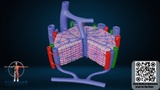
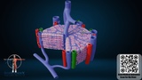
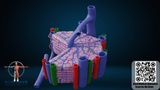
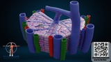
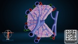
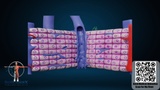
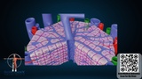
This Microscopic Anatomy of the Liver 3D Model illustrates the internal histological structure of the liver, focusing on the hepatic lobule, the fundamental functional unit of the liver.
The model visualizes important components such as hepatocytes, hepatic sinusoids, central vein, and portal triad, helping to demonstrate how blood flows through the liver and how metabolic processes occur within the organ.
This detailed scientific visualization is ideal for medical animation, histology education, biology teaching, and healthcare presentations. It provides a clear understanding of liver microstructure and the organization of liver tissue.
The model is suitable for anatomy training, medical research visualization, and educational explainer videos.