Descrição
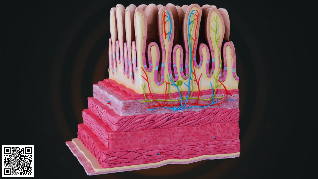
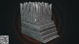
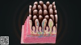
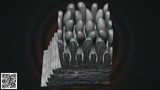
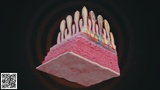
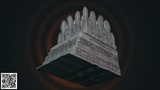
This highly detailed and anatomically accurate Human Small Intestine Histology 3D Model presents a comprehensive cross-sectional view of the intestinal wall, showcasing the complex structural organization responsible for digestion and nutrient absorption.
The model illustrates the inner mucosal surface lined with numerous finger-like projections called villi, which significantly increase the absorptive surface area of the small intestine. Between these villi, deep invaginations known as intestinal glands (Crypts of Lieberkühn) are clearly represented, highlighting the regenerative and secretory functions of the intestinal lining.
Each villus contains:
Rich capillary network (arterial and venous supply)
Central lacteal (lymphatic vessel for fat absorption)
Connective tissue core (lamina propria)
Simple columnar epithelial lining
Goblet cells for mucus secretion
The model also demonstrates the complete intestinal wall layering:
Mucosa – Includes epithelium, lamina propria, and muscularis mucosae
Submucosa – Contains blood vessels, lymphatics, and nerve plexus
Muscularis externa – Circular and longitudinal smooth muscle layers responsible for peristalsis
Serosa / outer connective tissue layer
The red and blue vascular structures depict oxygenated and deoxygenated blood circulation, while green structures represent lymphatic drainage. The muscular layers are distinctly separated, providing a clear visualization of intestinal motility mechanics.
The structural detailing makes this model suitable for microscopic-level educational visualization and professional medical presentations.